
sabato 22 dicembre 2012

martedì 18 dicembre 2012
UTILITA' DEGLI U.S. NEGLI ACCESSI VASCOLARI PERIFERICI
Studio che dimostra, ce ne fosse ancora il bisogno, il vantaggio nell'utilizzo degli u.s. negli accessi vascolari periferici "difficili"
J Nurs Care Qual. 2012 Jan-Mar;27(1):51-5. doi: 10.1097/NCQ.0b013e31822b4537.
Use of ultrasound guidance for peripheral intravenous placement in difficult-to-access patients: advancing practice with evidence.
Source
Louis A. Johnson VA Medical Center, Clarksburg, West Virginia 26301, USA. gmaiocco@hsc.wvu.edu
Abstract
Placement of peripheral intravenous (PIV) lines in difficult-to-access patients can be daunting. Multiple unsuccessful peripheral sticks, numerous PIV restarts, and potentially excess use of peripherally inserted central catheters can result. The goals of this project were to decrease the number of peripherally inserted central catheter referrals and lower the number of PIV restarts by having clinical nurses employ ultrasound guidance when initiating deep PIVs. After 10 months of nurses using the ultrasound as needed to insert a PIV line, the number of total peripherally inserted central catheter referrals decreased by 20%.
- PMID:
- 21826027
- [PubMed - indexed for MEDLINE]
UTILIZZO DELLA SAFENA IN PEDIATRIA
Pediatr Emerg Care. 2011 Dec;27(12):1121-5. doi: 10.1097/PEC.0b013e31823ab926.
Remember the saphenous: ultrasound evaluation and intravenous site selection of peripheral veins in young children.
Source
Pediatric Emergency Medicine, Yale-New Haven Children's Hospital, New Haven, CT 06504, USA. Antonio.riera@yale.edu
Proposta di approfondimento nel prendere in considerazione l'utilizzo della safena nei bambini al di sotto dei 3 anni
Abstract
OBJECTIVES:
The primary objective of this study was to measure the width and depth of peripheral veins using bedside ultrasound in children younger than 3 years. Secondary objectives included the evaluation of other vein and patient characteristics that may affect intravenous (IV) site selection. Assessment of nursing preferences for peripheral IV site selection was performed.
METHODS:
Sixty children aged 0 to 3 years who presented to an urban pediatric emergency department were enrolled. Ultrasound measurements of the transverse diameter (width) and distance from the top of the vein to the skin (depth) were recorded. Upon examination, veins were categorized as visible, palpable, detectable only by ultrasound, or not detectable. Sixteen staff nurses rated the likelihood of successful IV placement among different peripheral veins.
RESULTS:
The mean width of saphenous veins was significantly larger than that of hand veins (2.8 vs 1.8 mm, P < 0.0001). When comparing saphenous veins to antecubital veins, no significant difference was measured between the mean width (2.8 vs 2.8 mm). The mean depth of saphenous veins was significantly greater than those of hand veins (1.9 vs 1.4 mm, P < 0.0001) and antecubital veins (1.9 vs 1.6 mm, P = 0.019). Differences in visibility and palpability were observed between different vein types. Hand veins and antecubital veins were rated by the nursing staff as the most likely sites for successful IV placement, whereas saphenous veins were among the least likely (P < 0.0001).
CONCLUSIONS:
In children younger than 3 years, the saphenous vein is larger than hand veins and is similar in size to antecubital veins, although marginal differences in depth exist. The sonographic findings of the saphenous vein and antecubital vein suggest that either should be considered a superior first choice for IV cannulation in this age group. Knowledge of these differences is important when choosing a site for peripheral IV placement. Future studies should evaluate peripheral IV success rates by vein type with or without ultrasound guidance.
- PMID:
- 22134232
- [PubMed - indexed for MEDLINE]
lunedì 17 dicembre 2012
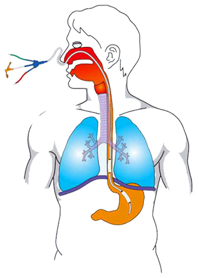
ACCESSI VENOSI IN NEONATOLOGIA
Report descrittivo su una complicanza causata da uno stravaso in caso di incannulamento di una vena epicranica in un neonato.
Non è strettamente pertinente all'ecografia..........forse.......
Anche se è solo un caso si può evincere che le complicanze possono essere anche serie e una domanda ai colleghi che operano con i piccoli: a voi è mai capitato? (anche se non di questa entità).
Buona lettura
Adv Neonatal Care. 2011 Aug;11(4):251-4. doi: 10.1097/ANC.0b013e31822565b4.
Preterm infant with subdural hematoma from malpositioned scalp intravenous catheter.
Source
Department of Radiology, Medical University of South Carolina, Charleston, SC 29425, USA. Meanss@musc.edu
Abstract
Vascular access is critical in the care of sick infants and children for the direct administration of medications and fluids. In infants, especially preterm infants, the use of scalp veins is a common practice because of less subcutaneous fat and less mobility around the catheter site decreasing the risk of dislodgement. We describe a case of a 24-week preterm infant girl born via caesarean section delivery who developed signs of increased intracranial pressure on day of life 11. A head computed tomography (CT) demonstrated large bilateral subdural hematomas with midline shift secondary to packed red blood cell infusion via an incorrectly positioned scalp intravenous catheter in the subdural space. In general, the use of scalp veins for intravenous access is a common method for direct administration of medications and fluids in small infants, with risks that are comparable to those associated with peripheral venous access. The use of scalp intravenous catheters is a fairly safe practice when correctly positioned. Position confirmation before and during use is vital to avoid potential intracranial complications.
- PMID:
- 22123346
- [PubMed - indexed for MEDLINE]
venerdì 14 dicembre 2012
ARTERIA RADIALE
Qui di seguito riporto un articolo di Chest sul cateterismo dell' Arteria Radiale in Ecoguida
Chest. 2011 Mar;139(3):524-9. Epub 2010 Aug 19.
Ultrasound-guided catheterization of the radial artery: a systematic review and meta-analysis of randomized controlled trials.
Source
Division of Critical Care Medicine, Montefiore Medical Center and The Albert Einstein College of Medicine, 111 E 210th St, Bronx, NY 10467, USA. arielshiloh@gmail.com
Abstract
BACKGROUND:
Ultrasound guidance commonly is used for the placement of central venous catheters (CVCs). The Agency for Healthcare Research and Quality recommends the use of ultrasound for CVC placement as one of its 11 practices to improve patient care. Despite increased access to portable ultrasound machines and comfort with ultrasound-guided CVC access, fewer clinicians are familiar with ultrasound-guided techniques of arterial catheterization. The goal of this systematic review and meta-analysis was to determine the utility of real-time two-dimensional ultrasound guidance for radial artery catheterization.
METHODS:
A comprehensive literature search of Medline, Excerpta Medica Database, and the Cochrane Central Register of Controlled Trials by two independent reviewers identified prospective, randomized controlled trials comparing ultrasound guidance with traditional palpation techniques of radial artery catheterization. Data were extracted on study design, study size, operator and patient characteristics, and the rate of first-attempt success. A meta-analysis was constructed to analyze the data.
RESULTS:
Four trials with a total of 311 subjects were included in the review, with 152 subjects included in the palpation group and 159 in the ultrasound-guided group. Compared with the palpation method, ultrasound guidance for arterial catheterization was associated with a 71% improvement in the likelihood of first-attempt success (relative risk, 1.71; 95% CI, 1.25-2.32).
CONCLUSIONS:
The use of real-time two-dimensional ultrasound guidance for radial artery catheterization improved first-pass success rate.
- PMID:
- 20724734
- [PubMed - indexed for MEDLINE]
domenica 9 dicembre 2012
VERIFICA POSIZIONE DEL S.N.G. ATTRAVERSO L'ECOGRAFO
 Un articolo tratto dallo Scandinavian Journal of Trauma, Resuscitation and Emergency Medicine che tratta di uno studio che compara l'auscultazione classica in epigastrio Vs l'utilizzo dell'ecografo per valutare il corretto posizionamento del sondino naso gastrico.
BUONA LETTURA!
http://www.sjtrem.com/content/pdf/1757-7241-20-38.pdf
giovedì 6 dicembre 2012
SKI LIFT
TRATTO DA: http://resusme.em.extrememember.com/category/ultrasound/page/4/
“Ski Lift” Ultrasound Technique
In Dr Jason Nomura’s excellent blog takeokun.com he describes his technique to assist in viewing the needle during in-plane ultrasound guidance for vascular access.
The technique is described as follows:
- Obtain a sagittal view of the target vessel
- Stabilize the transducer and brace your hand. Then rock the probe to elevate the proximal section.
- Place the needle in the center of the probe (usually at the case seam) and under the probe footprint.
- Stop rocking the probe so the entire surface is again contacting the skin, the needle tip should be immediately visible.
- Advance the needle to the target vessel
Click the image below to go to the site and the demonstration video:

The “Ski Lift”: A Technique to Maximize Needle Visualization with the Long-axis Approach for Ultrasound-guided Vascular Access
Acad Emerg Med. 2010 Jul;17(7):e83-4
Ho seguito il consiglio del collega Romei ed effettivamente grossi miglioramenti non ne ho visti rispetto alle consuete tecniche.... vedremo di aumentare la manualità...... vediamo in seguito....
Altra domanda: l'asepsi in caso di incanulamenti di vene centrali? Non c'è un rischio di "bucare" la guaina protettiva sterile della sonda con il rischio di inquinare il campo?
Mi fate sapere se vi siete trovati bene con questa tecnica?
martedì 4 dicembre 2012
CORSO DI FORMAZIONE TORINO
Annuncio della replica del corso tenutosi l'anno scorso .
Appena possibile vi descriverò le modalità d'iscrizione e gli indirizzi utili.
Il costo dovrebbe essere circa quello dell'anno scorso più o meno 100 euro
UNIVERSITÀ DEGLI STUDI DI TORINO
corso in ECOGRAFIA il 22-23-24 maggio 2013
Istituto Rosmini di Torino
Provider: Università degli Studi di Torino - (ID 173)
Accr. 173-30203. I
crediti attribuiti= 29,5.
IMPIEGO DEGLI ULTRASUONI NELL’ASSISTENZA INFERMIERISTICA ED OSTETRICA: ECOGRAFIA DI SUPPORTO E CONTROLLI DI QUALITÀ
Istituto Rosmini - Aula _Magna_- Via Rosmini,
1° GIORNATA: __22 maggio 2013
Dalle ore 08:30 alle ore 10:30 Titolo/argomento Docenti
Dalle ore 8.30 alle ore 9.00 Saluto delle Autorità
Rappresentanti dell’Università e degli ordini professionali
Dalle ore 9.00 alle ore 9.45 Stato dell’arte
G. Cibinel
A.Prone
Dalle ore 9.45..alle ore 10.30 Buone pratiche nell’utilizzo dell’ecografo: l’esperienza SIEOG T. Todros
Dalle ore 11:00 alle ore 13:00 Titolo/argomento Docenti
Dalle ore 11.00..alle ore 11.45 Principi di fisica degli ultrasuoni e formazione dell’immagine ultrasonografica.
R. Spagnolo
Dalle ore 11.45.alle ore 12.30. Problematiche generali di Controllo di Qualità e loro correlazione con i rischi per professionisti e utenti C. Guiot
Dalle ore 12.30.alle ore 13.00. Confronto Dibattito
Dalle ore 14:00 alle ore 18:00 ASPETTI GIURIDICO-LEGALI ED ECONOMICO-ORGANIZZATIVI
Docenti
Dalle ore 14 alle ore 15.30 L’uso dell’ecografia di supporto nell’assistenza ostetrica ed infermieristica P. Serafini
C. Mesiti
G. Borasi
Dalle ore 15.30 alle ore 16.15 Profili di responsabilità nella metodica ultrasonografica G. Marzo
Dalle ore 16.15 alle ore 17.00 Ultrasonologia: aspetti economici e organizzativi" N. Dirindin
Dalle ore 17.00 alle ore 17.45 Laboratori di Ultrasonologia e problematiche di certificazione E. Dragone
Dalle ore 17.45 alle ore 18.00 Confronto Dibattito
2° GIORNATA: __23 maggio 2013
Dalle ore 08:45 alle ore 10:30 ULTRASONOLOGIA IN AMBITO INFERMIERISTICO ED OSTETRICO
Docenti
Dalle ore 8.45 alle ore 9.30 Tecniche di imaging sonografico dei vasi arteriosi e venosi P.Pasquero
Dalle ore 9.30 alle ore 10.30 Imaging ecografico nell’impianto di cateteri ad inserzione periferica :
esperienze e prospettive
A. Bettineschi
G. Borasi
Dalle ore 11:00alle ore 13:00 Titolo/argomento Docenti
Dalle ore 11.00 alle ore 11.45 Complicanze nell’ impianto di cateteri centrali e periferici
S. Sandrucci
Dalle ore 11.45 alle ore 12.45 Imaging ecografico in emergenza generale, pediatrica ed ostetrica A. Urbino,
E. Viora
Dalle ore 12.45 alle ore 13.00 Confronto Dibattito
Dalle ore 14:00 alle ore 17:30 Titolo/argomento Docenti
Dalle ore 14.00 alle ore 15.00 Tavola rotonda: utilizzo dell’ecografia nella visualizzazione della vescica in ambito pediatrico, infermieristico e ostetrico-ginecologico
L. Romei,
M. Catti,
L. Priori
Dalle ore 15.00 alle ore 17.45 Esercitazioni in laboratorio con apparecchiature ultrasonografiche ed utilizzo di phantoms
(c/o Ist Rosmini) L. Canavese,
C. Guiot,
L. Merlone,
P. Pasquero,
G. C. Musso
test di apprendimento delle prime due giornate.
TERZA GIORNATA: 24 maggio 2013
Attività Pratica :
le nozioni apprese vengono applicate nell’ambito delle applicazioni professionali specifiche. Si rende pertanto necessaria una suddivisione tra ostetriche e infermieri e l’utilizzo di ecografi dedicati da utilizzare in ambito clinico su pazienti e/o volontari
.
ore 8:30 ore 12:30 Titolo/argomento Docenti
Gruppo A +B
Ospedale S. Anna Introduzione alla esecuzione della ecografia in gravidanza, indicazioni e finalità nelle varie epoche.
Dall’informazione basata su prove di efficacia alla scelta informata delle donne, ruolo dell’ostetrica.
Principi e tecniche di base per l’ecografia di supporto
Esercitazioni pratiche e conduzione dell’indagine ultrasonografica su paziente: gruppi di tre discenti per apparecchiatura ultrasonografica
Verrà svolto in modalità comune tra gruppo A (che nel pomeriggio proseguirà all’ osp S. Anna) e gruppo B (che nel pomeriggio proseguirà all’ osp. M. Vittoria) P.Gaglioti
L. Priori
E.Viora
A. Sciarrone
M.Canesi
L.Canavese,
B. Contino
S.Cantoira
Gruppo C infermieri
Istituto Rosmini
Esercitazioni pratiche e conduzione dell’indagine ultrasonografica su paziente:
ecografia topografica del distretto vascolare dell’arto superiore su volontario sano
utilizzo dell’ecografia come supporto nel reperimento di accessi vascolari, esercitazioni pratiche su fantoccio
l’utilizzo della metodica ultrasonografica nel monitoraggio della vena cava inferiore come indice di “riempimento” del paziente
lo studio vescicale ed il calcolo della volumetria in volontari sani
cenni di ecografia polmonare, utilità in triage
L. Merlone
G. Borasi
P. Pasquero
C. Mesiti
A. Palumbo
A. Bettineschi
ore 13:30 ore 18:00 Titolo/argomento Docenti
Gruppo A
Ospedale S. Anna
Esercitazioni pratiche e conduzione dell’indagine ultrasonografica su paziente: gruppi di tre discenti per apparecchiatura ultrasonografica su casi selezionati per la rilevazione della presentazione della p.p., attività cardiaca fetale, A.F.I., localizzazione della placenta, etc.
P.Gaglioti
L. Priori
E.Viora
A. Sciarrone
M.Canesi
Gruppo B
Ospedale Maria Vittoria
Esercitazioni pratiche e conduzione dell’indagine ultrasonografica su paziente: gruppi di tre discenti per apparecchiatura ultrasonografica su casi selezionati per la rilevazione della presentazione della p.p., attività cardiaca fetale, A.F.I., localizzazione della placenta, etc.
L.Canavese
B. Contino
S.Cantoira
Gruppo C infermieri
Istituto Rosmini
Esercitazioni pratiche e conduzione dell’indagine ultrasonografica su paziente: gruppi di tre discenti per apparecchiatura ultrasonografica. Completamento e verifica del gruppo C della sessione mattutina
ecografia topografica del distretto vascolare dell’arto superiore su volontario sano
utilizzo dell’ecografia come supporto nel reperimento di accessi vascolari, esercitazioni pratiche su fantoccio
l’utilizzo della metodica ultrasonografica nel monitoraggio della vena cava inferiore come indice di “riempimento” del paziente
lo studio vescicale ed il calcolo della volumetria in volontari sani
cenni di ecografia polmonare, utilità in triage
L. Merlone
G. Borasi
P. Pasquero
C. Mesiti
A. Palumbo
A. Bettineschi
VALUTAZIONE finale
Il tempo dedicato alla verifica NON è compreso nelle ore totali del corso
TOTALE ORE DELL’INIZIATIVA FORMATIVA 24
ORE INTERATTIVE 11.15
sabato 1 dicembre 2012
eeeeh gli infermieri!!!
dal sito: http://resusme.em.extrememember.com/category/ultrasound/
Dallo stesso sito un bell'articolo Lung ultrasound for pneumothorax by paramedics che vi posto qui di seguito....BUONA LETTURA!!!
Lung ultrasound for pneumothorax by paramedics
Comments Off
 This UK study showed that paramedics could successfully acquire and identify lung ultrasound images after a two day course. The course covered the identification and management of patients who present with serious thoracic injury, with a specific focus on the use of thoracic ultrasound during early prehospital assessment. Standard 2D images for pleural sliding and comet tails and M-Mode for the ‘seashore sign’ were acquired, and colour Doppler was also used to assist in the identification of pleural sliding.
Objective This trial investigated whether advanced paramedics from a UK regional ambulance service have the ability to acquire and interpret diagnostic quality ultrasound images following a 2-day programme of education and training covering the fundamental aspects of lung ultrasound.
Method The participants were tested using a two-part examination; assessing both their theoretical understanding of image interpretation and their practical ability to acquire diagnostic quality ultrasound images. The results obtained were subsequently compared with those obtained from expert physician sonographers.
Results The advanced paramedics demonstrated an overall accuracy in identifying the presence or absence of pneumothorax in M-mode clips of 0.94 (CI 0.86 to 0.99), compared with the experts who achieved 0.93 (CI 0.67 to 1.0). In two-dimensional mode, the advanced paramedics demonstrated an overall accuracy of 0.78 (CI 0.72 to 0.83), compared with the experts who achieved 0.76 (CI 0.62 to 0.86). In total, the advanced paramedics demonstrated an overall accuracy at identifying the presence or absence of pneumothorax in prerecorded video clip images of 0.82 (CI 0.77 to 0.86), in comparison
with the expert users of 0.80 (CI 0.68 to 0.88). All of the advanced paramedics passed the objective structured clinical examination and achieved a practical standard considered by the examiners to be equivalent to that which would be expected from candidates enrolled on the thoracic module of the College of Emergency Medicine level 2 ultrasound programme.
Conclusion This trial demonstrated that ultrasound-naive practitioners can achieve an acceptable standard of competency in a simulated environment in a relatively short period of time.
Acquisition and interpretation of focused diagnostic ultrasound images by ultrasound-naive advanced paramedics: trialling a PHUS education programme
Emerg Med J, 2012 vol. 29 (4) pp. 322-326
Emerg Med J, 2012 vol. 29 (4) pp. 322-326
Iscriviti a:
Post (Atom)